




Case 1
“They Said It Couldn’t be Done…”
By Dr. Kirk Noraian
On April 19, 2011, the patient, a 47 year-old Caucasian female, presented with the chief complaint on tooth #31 of “I need bone added to this tooth.” The referral card received from the referring dentist indicated “#31 Bone loss. Needs bone added.” In accordance with our office policy to provide a comprehensive examination, a full mouth periodontal charting was completed, and an intra-oral examination with oral pathology screening was performed. Radiographs were not immediately available for review. A blue lesion 2cm in diameter was noted on the right lateral border and dorsum of the tongue. The patient had this lesion for many years and had been told by several physicians that the lesion is inoperable. The patient presented with probing depths as deep as 8mm and occlusal prematurities were noted. Radiographic evidence of advanced bone loss was noted.
The diagnoses were as follows: 1) Generalized Advanced (Type IV) Chronic Periodontitis pending radiographic confirmation; 2) Generalized occlusal prematurities; and 3) Hemangioma of Tongue.
Pending confirmation with radiographs, the patient was offered the following treatment options: No Treatment; Periodontal Maintenance; Scaling and Root Planing; Guided Bone Regeneration; Laser Assisted New Attachment Procedure (LANAP); and laser ablation of the tongue lesion with the PerioLase MVP-7 Nd:YAG laser. Aware of the risks, benefits and alternatives to treatment, the patient elected to proceed with the LANAP pending radiographic confirmation and laser ablation of the tongue lesion with the PerioLase MVP-7 Nd:YAG laser. The patient was informed that the goal of laser treatment is to use the least amount of energy possible so as not to cause irreparable harm to the tissue, therefore, multiple visits may be required to completely remove the lesion.
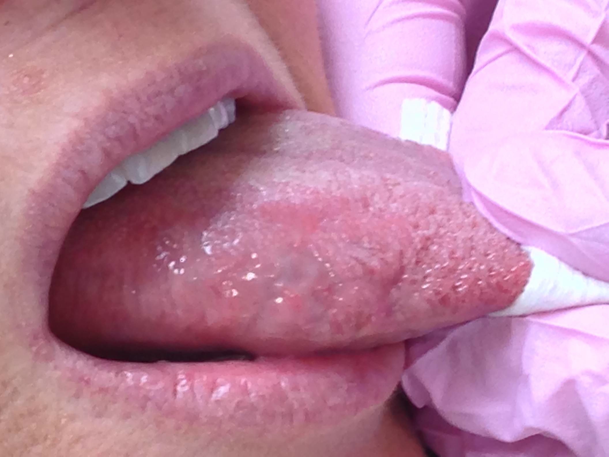
On April 16, 2012, the patient returned for surgery as planned and radiographic confirmation was received of her periodontal status. The patient presented without complaint. The lesion had a bluish color with a central red area and measured about 2cm in diameter. (Figure 1). The patient received moderate sedation in our usual manner for full mouth surgery. Local anesthetic was administered via bilateral inferior alveolar, long buccal and mental blocks with buccal and lingual infiltration with aspirating as would be common for the LANAP procedure and provide the secondary benefit of anesthetizing the tongue. Direct injection of the lesion was avoided given vascularity of the tongue. Once the LANAP was completed, the patient’s tongue was addressed with ablation using the PerioLase MVP-7 Nd:YAG laser on the Laser Hemostasis Setting in the following manner: Pulse Duration: 650usec; Energy per pulse: 200mJ; Repetition Rate: 20Hz; Average Power: 4.0W; with a Total Energy: 1819J. The reason for selecting the Laser Hemostasis Setting is the advantage of deeper energy penetration of the tissue. The lesion was treated with the laser not in contact with the tissue to get a feel at first of how the lesion would react to the energy administered. The best description for what was observed would be like slowly letting the air out of a tire. It was obvious the central, deeper core of the lesion was going to be more difficult to treat, at which time the optic fiber was introduced into the tissue. The lesion shows evidence of a white sloughing appearance with a blue core where the lesion was deepest. Figure 2.
On April 17,2012, the patient returned for a 24 hour post-operative visit with a chief complaint “I am afraid the Peridex will burn my tongue.” Tongue lesion appeared white in color, and a dark spot was evident where Hemangioma was the deepest. Soft tissue had started to slough about the now white lesions and the tongue appeared slightly swollen. (Figure 3).
On April 23, 1012, the patient returned for a one week post-operative visit with a chief complaint of “It feels like a piece is going to fall off.” The sloughing is more pronounced about the lesion as the initial white layer was off and a deeper yellow sloughing tissue was noted. The swelling of the tongue was not as pronounced. (Figure 4).
On April 30,2012, the patient returned for two week post-operative visit with the chief complaint “I’m glad my tongue isn’t so swollen anymore.” The tongue wound had a red and white appearance with sloughing that appeared to be resolving. The dark central core of the lesion was evident still. (Figure 5).
On May 14, 2012, the patient returned for a one month post-operative visit with the chief complaint of “My right side just feels different but not uncomfortable.” The borders of the lesion have started to turn pink with the reddish-blue core measuring about 9mm in diameter. Clearly the lesion was getting smaller. (Figure 6).
On June 21, 2012, the patient returned for a nine week post-operative visit with the chief complaint of “We need to do something about this,” referring to the protruding lesion from her tongue. The lesion is sessile with narrow stalk. (Figures 7 & 8). The appearance could be described as though the firmer adjacent tongue tissue was squeezing out the vascular lesion. The patient agreed to return for excisional biopsy of the lesion. The return for additional surgery was not a problem for the patient as she was aware from the beginning that retreatment may be indicated.
On June 26, 2012, the patient returned to have the lesional tissue excised and had a chief complaint “I am so ready for this to come off.” At this point, the patient had a persistent cavernous hemangioma. Aware of the risks, benefits, and alternatives, the patient agreed to proceed with excisional biopsy on this date. Local anesthetic was administered with inferior alveolar block, intralingual and intralesional infiltration, while using aspirating technique. First, the PerioLase MVP-7 Nd:YAG Laser was used on Diode Mode Setting in the following manner: Pulse Duration: 100usec, Energy per Pulse: 100mJ, Repetition Rate: 100Hz, Average Power: 4.0W, with Total Energy: 404J. to excise the lesion. The settings were changed to the Gingivectomy Setting to clean up the tissue edges in the following manner: Pulse Duration: 100usec,, Energy per Pulse: 180mJ, Repetition Rate: 20Hz, Average Power: 3.6W with a Total Energy: 105J. The combined energy for both steps was 509J., The excised lesional tissue of about 10mm in diameter was placed in a commonly used formaldehyde solution and forwarded to the Surgical Pathology Service at the University of Kentucky and on the requisition slip it was noted that the excisional biopsy was performed with a laser. (Figures 9 & 10).
On June 27, 2012, the patient returned for a 24 hour post-operative visit without complaint. The lesion is sloughing and an indentation is noted in the soft tissue from where the biopsy was excised. (Figure 11).
On July 2,2012, the patient returned for a one week post-operative visit with a chief complaint of “I hardly had any pain at all.” The sloughing is more evident and the diameter of the tissue defect is decreasing.
On July 16, 2012, the patient returned for a three week post-operative visit with a chief complaint of “I feel good.” The sloughing is gone and a small dimple is evident in the soft tissue from where the excisional biopsy was taken. (Figure 13).
On July 3, 2012, the Surgical Pathology Report is received from the University of Kentucky with a confirming diagnosis consistent with a Cavernous Hemangioma. The patient is notified of this report.
On July 17, 2013, the patient returns about one year later because she noticed the lesion was not completely obliterated and she has the following chief complaint, “I came in because I thought I could get it taken care of. We talked about it the last time and I wanted you to see it. I wanted to return so others could see how this is treated. I have no pain.” The lesion appeared bluish characteristic of the underlying venous vasculature. (Figure 14). The patient demonstrates a persistent Cavernous Hemangioma. Aware of the risks, benefits, and alternatives to treatment, the patient elected to proceed with another ablative procedure with the understanding that additional treatments may be required. Using the PerioLase MVP-7 Nd:YAG laser on the LPT Hemostasis Setting not in contact as follows: Pulse Duration: 650usec, Energy per Pulse: 200mJ, Repetition Rate: 20Hz, Average Power: 4.0W with a Total Energy: 862J. The now smaller lesion underwent a round of “deflation” similar to what was noted during the first procedure. The lesion has a white appearance consistent with early sloughing. (Figure 15).
On July 18, 2012, the patient returned for a 24 hour post-operative visit with a chief complaint of “It is sore, but it’s ok. There was some swelling this morning but seems better now.” The sloughing is noted as well as some exposure of the underlying connective tissue. (Figure 16).
On July 31, 2013, the patient returned for a two week post-operative visit and had a chief complaint, “It is better now.” Two dimples are noted in the healing soft tissue. The one where the deeper portion of the lesion was noted, clearly demonstrated deeper dimpling. (Figure 17).
On September 10, 2013, the patient returned for a two month post-operative visit with the chief complaint of “It feels pretty good.” The dimples appeared smoother and some vasculature of the lesional tissue was evident. (Figure 18). The patient was informed that additional retreatment of the hemangioma is likely and more prompt attention would likely help reduce the vascular lesion before the vessels have a chance to extravasate and outpouch.
On November 14, 2012, the patient returned for her periodontal maintenance visit without complaint. The tissue appears to be maturing as expected and the lesional tissue is still evident. The patient would like to proceed with retreatment after the holidays and she will get back to us when she is ready to proceed.
Figure 1. Pre-Operative.
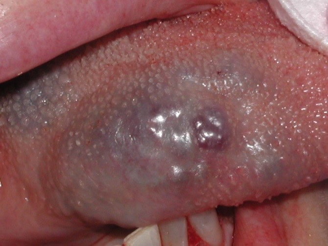
Figure 2. Immediate Post-Operative.
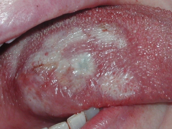
Figure 3. 24 Hours Post-Operative.
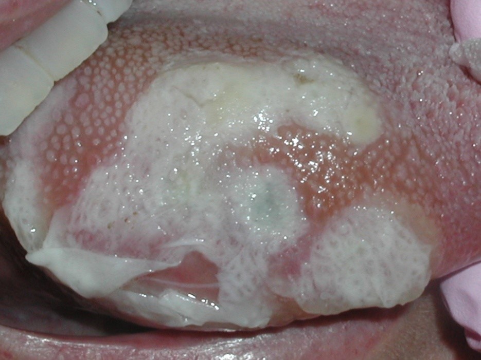
Figure 4. 1 Week Post-Operative.
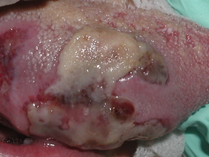
Figure 5. 2 Weeks Post-Operative.
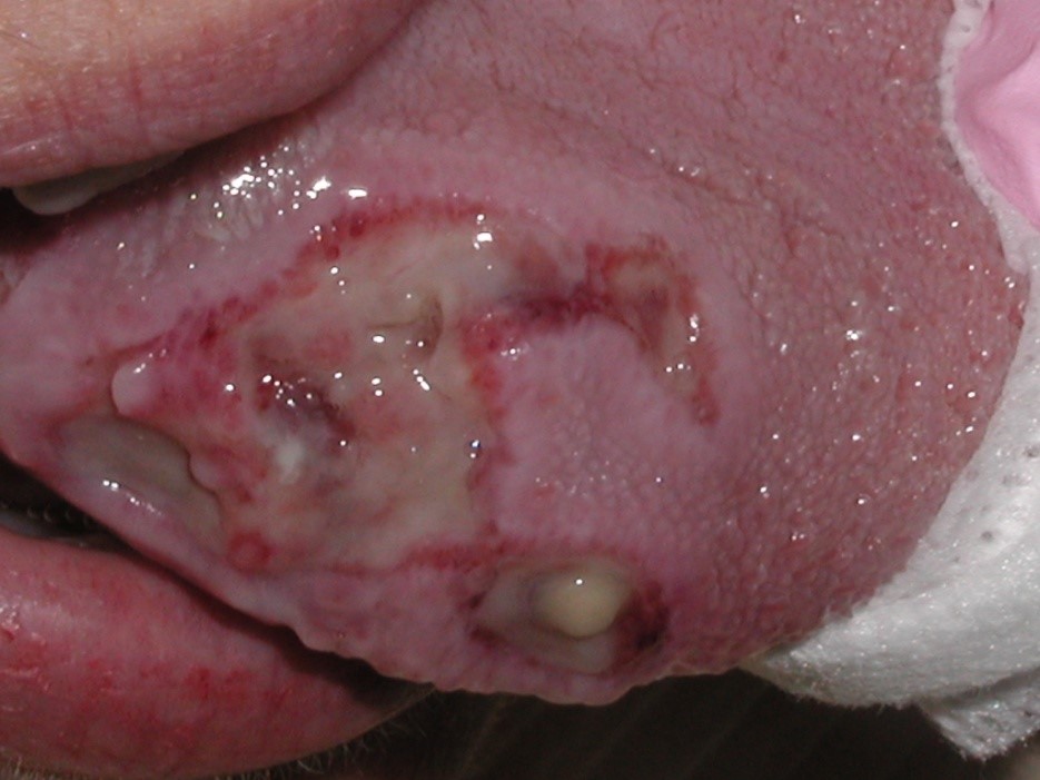
Figure 6. 4 Weeks Post-Operative.
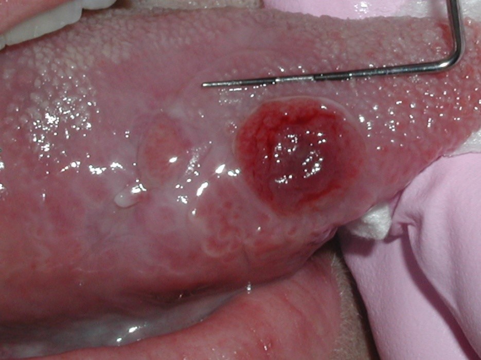
Figure 7. 9 Weeks Post-Operative.
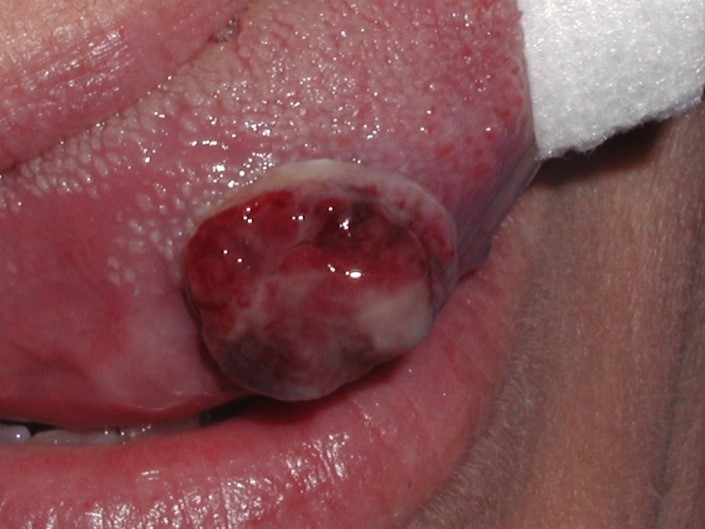
Figure 8. 9 Weeks Post-Operative demonstrating sessile nature.
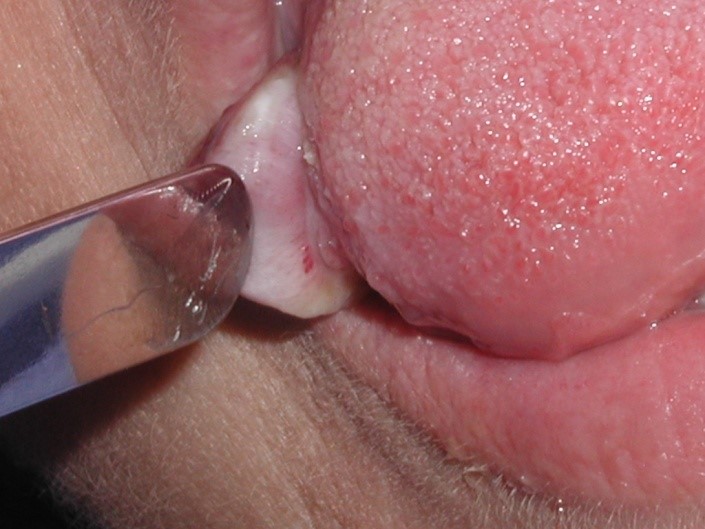
Figure 9. Excisional Biopsy Immediately Post-Operative.
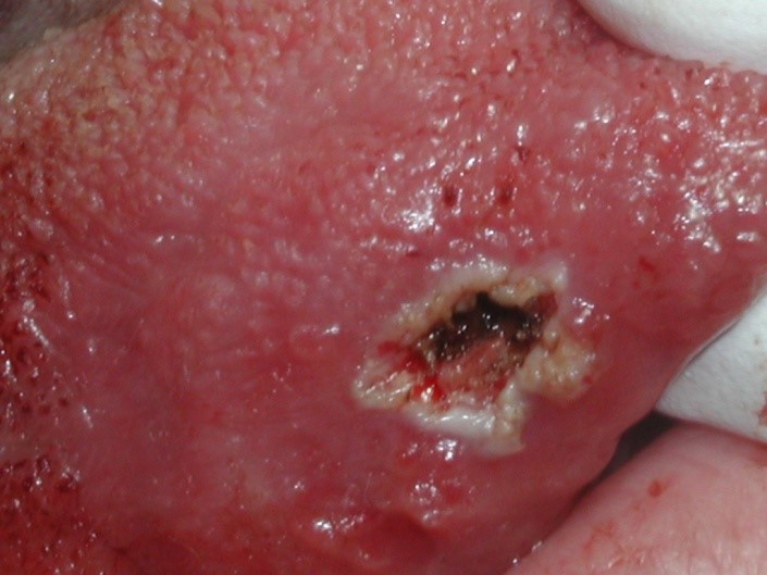
Figure 10. Excised lesional tissue.
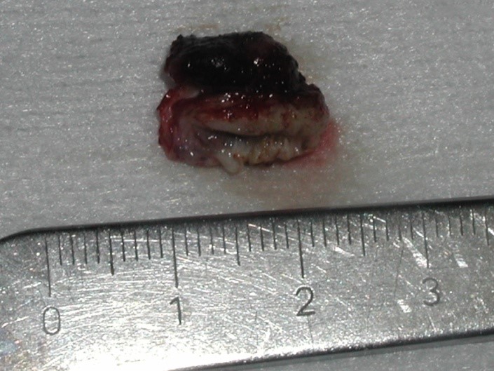
Figure 11. Excisional Biopsy 24 Hours Post-Operative.
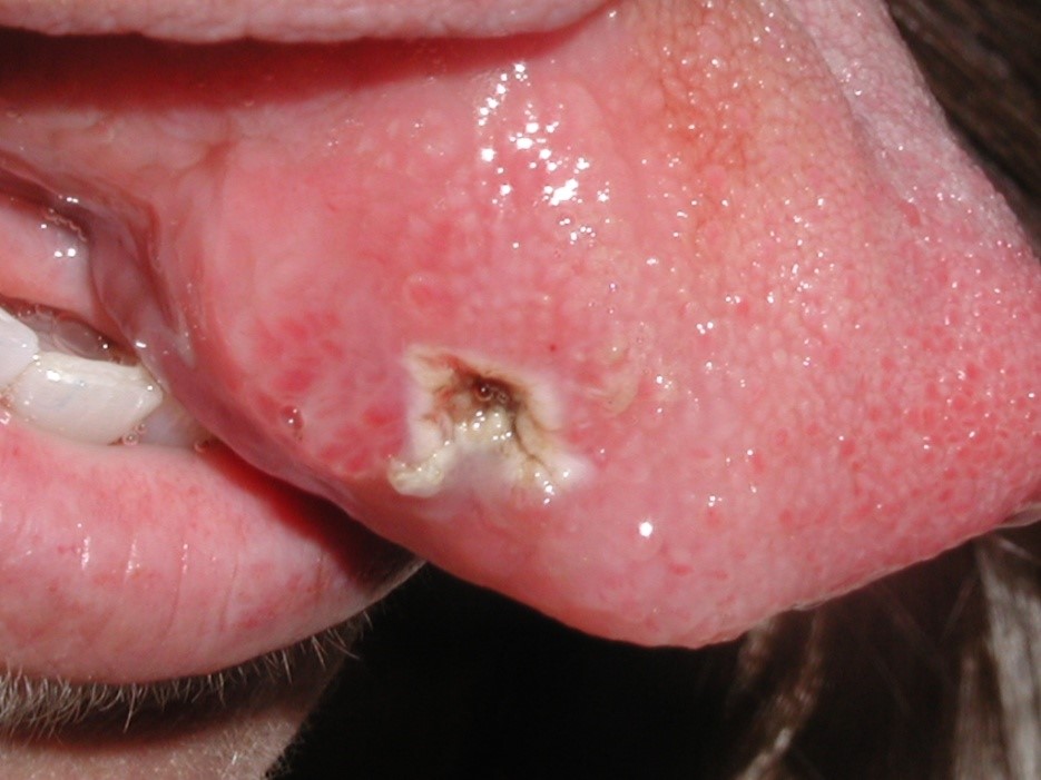
Figure 12. Excisional Biopsy – 1 Week Post-Operative.
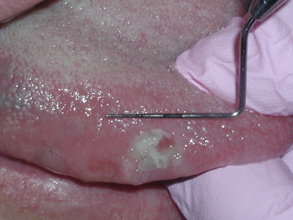
Figure 13. Excisional Biopsy – 3 Weeks Post-Operative.
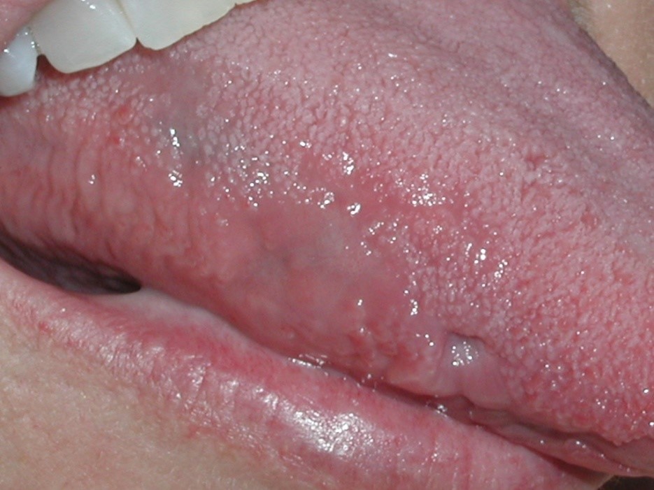
Figure 14. Vasculature apparent in recurrent lesion.
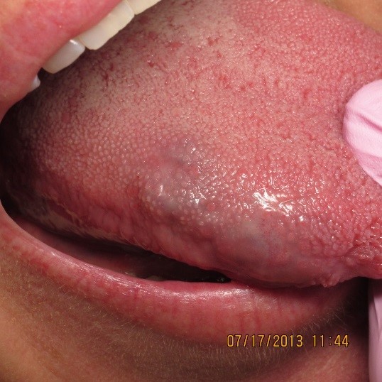
Figure 15. 3rd Procedure – Immediately Post-Operative.
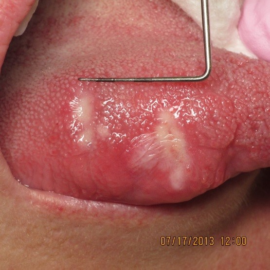
Figure 16. 3rd Procedure – 24 Hours Post-Operative.
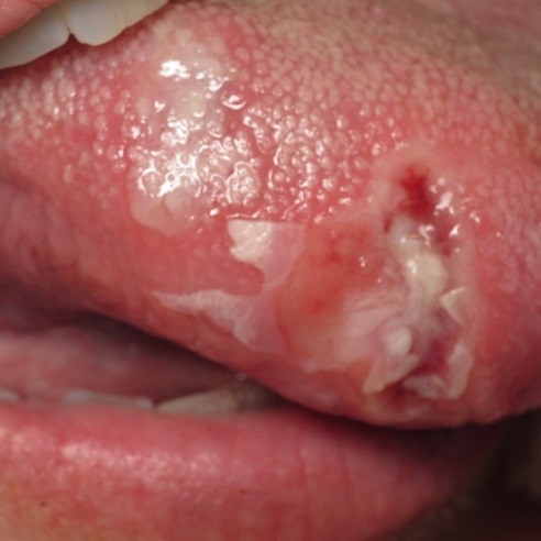
Figure 17. 3rd Procedure – 2 Weeks Post-Operative.
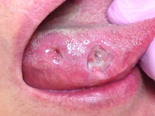
Figure 18. 3rd Procedure – 2 Months Post-Operative.
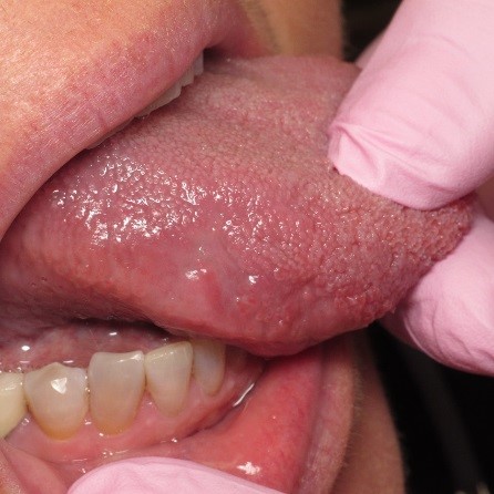
Figure 19. 3rd Procedure – 4 Months Post-Operative.
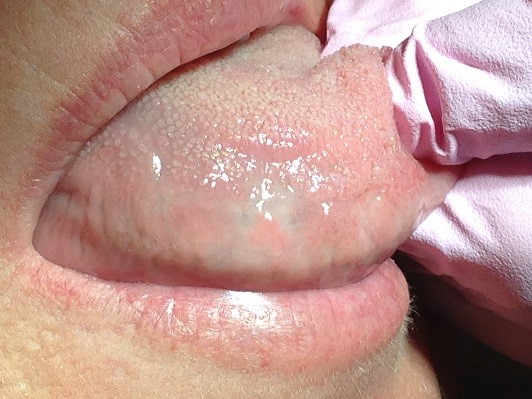
Final post-op